by tatkonasajt | Aug 7, 2023
This course is approved by GRSK and will cover the following subjects:
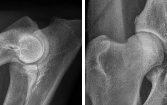
- Introduction to hip dysplasia and elbow dysplasia.
- Possibilities to reduce dysplasia: how can breeding influence the prevalence of HD and ED.
- Radiographic technique – practical on how to take a proper radiographs for the assessment of the hip and elbow dysplasia. Quality evaluation of the radiographs for the HD and ED.
- Explanation of different scoring systems.
- Film reading session – scoring of the HD ED radiographs.
DAY1
| Time |
Title |
| 09.00 – 09.15 |
Welcome and Introduction |
| 09.15 – 10.30 |
Introduction to hip dysplasia and elbow dysplasia
Possibilities to reduce dysplasia
How can breeding influence the prevalence of HD and ED |
| 10.30 – 11.00 |
Coffee break |
| 11.00 – 12.00 |
How to take a proper radiograph for the HD assessment |
| 12.00 – 13.00 |
How to take a proper radiograph for the ED assessment |
| 13.15 – 14.30 |
Lunch break |
| 14.30 – 16.30 |
Radiographic technique – practical |
| 16.30 – 17.00 |
Coffee break |
| 17.00 – 18.30 |
Film reading session on radiographic film quality |
DAY2
| Time |
Title |
| 09.00 – 10.30 |
HD scoring systems / protocols
Radiographic findings and evaluation of HD
Scoring criteria and grading of HD |
| 10.30 – 11.00 |
Coffee break |
| 11.00 – 12.30 |
HD film reading individual work and group work |
| 12.30 – 13.30 |
F.C.I. – Kennel Clubs, Breed Clubs – HD ED control |
| 13.30 – 14.30 |
Lunch break |
| 14.30 – 16.00 |
ED scoring system / protocol
Radiographic findings and evaluation of ED
Scoring criteria and grading of ED |
| 16.00 – 16.30 |
Coffee break |
| 16.30 – 18.00 |
ED film reading individual work and group work |
| 18.00 – 18.30 |
Closing remarks |
by tatkonasajt | Apr 17, 2023
DAY 1
- Clinical pharmacology of sedatives, analgesics and hypnotics in veterinary anaesthesia
- Clinical use of intraoperative monitoring and hemodynamic support
- Cardiovascular complications
- Respiratory complications
DAY 2
- Other complications during anaesthesia
- Anaesthesia of a patient with heart disease
- Anaesthesia of a patient with liver/kidney disease
- Anaesthesia of patients with respiratory/neurological/endocrine disease
by tatkonasajt | Mar 26, 2023
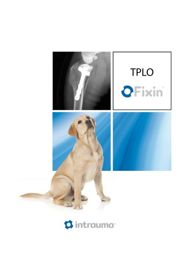
Objectives and goals of the course
This TPLO course is designed for practicing veterinarians with experience in small animal orthopaedic surgery. The participants will became familiar with the planning and execution of the most commonly performed procedure in cranial cruciate deficient stifle using one of the most versatile TPLO locking system.
The detailed theoretical sessions will bring the knowledge of rationale, case selection, preoperative planning, surgical execution and potential complications in TPLO procedure. The structure of the labs will allows the participants to learn the procedure in a “step by step” manner first on a bone models and then on a cadaver species.
DAY 1
| Time |
Title |
| 08:30 – 09:00 |
Understanding stifle biomechanics and rationale behind TPLO |
| 09:00 – 09:30 |
Radiographic positioning and TPA measuring |
| 09:30 – 10:00 |
TPLO implants and equipment |
| 10:00 – 10:30 |
Coffee break |
| 10:30 – 11:00 |
Preoperative planning of TPLO |
| 11:00 – 12:30 |
TPLO – step by step description of the procedure. Postoperative radiographic assessment after TPLO |
| 12:30 – 13:30 |
Lunch break |
| 13:30 – 14:00 |
Complications related to the TPLO procedure |
| 14:00 – 15:00 |
Preoperative planning of TPLO – practical exercise (drawing on a printed radiographs, TPA calculation, defining the best position of the cut, implant choice and position) – TPLO charts, saw blade radius templates and plates templates will be needed for this exercise |
| 15:00 – 17:00 |
Getting familiar with the equipment and performing TPLO on a bone model – laboratory |
| 17:00 – 17:30 |
Summary and question session and end of the course |
DAY 2
| Time |
Title |
| 08:30 – 10:00 |
Preoperative planning on x-rayed cadaver limbs |
| 10:00 – 10:30 |
Coffee break |
| 10:30 – 12:00 |
TPLO on a cadaver limb – practical session 1 |
| 12:00 – 13:00 |
Lunch break |
| 13:00 – 14:30 |
TPLO on a cadaver limb – practical session 2 |
| 14:30 – 15:30 |
Discussion of postoperative radiographs |
| 15:30 – 16:00 |
What’s the difference in Giant breeds |
| 16:00 – 16:30 |
What’s the difference in small dogs. TPLO + patella luxation |
| 16:30 – 17:00 |
Summary, question session and end of the course |
by tatkonasajt | Mar 26, 2023
Objectives and goals of the course
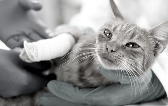
This two days course is designed for veterinary practitioners with some experience in treatment of fractures and other orthopaedic conditions in small animals. Some anatomical differences makes the orthopaedic care in feline patients more challenging and might reflects to the treatment options. The course will consists of theoretical sessions alternating with small group practicals on cadaver specimens. This will allows the participants to implement the knowledge delivered during the theoretical parts in the practical sessions starting with performing the surgical approach to different bones and using different implants for stabilising some of the most commonly seen fractures in feline patients.
Overview of most common fracture scenarios in feline patients
Decision making process in fracture planning
Interactive case-oriented discussion
After the course, participants will be able to construct a treatment plan and will become familiar how to treat most common orthopaedic conditions and fractures in feline patients.
DAY 1
| Time |
Title |
| 09:00 |
Welcome |
| 09:15 – 10:15 |
Principles of fracture repair in cats – decision making and treatment options |
| 10:15 – 11:15 |
Common fractures of the front limb |
| 11:15 – 11:30 |
Coffee break |
| 11:30 – 12:30 |
Common fractures of the hind limb |
| 12:30 – 13:30 |
Lunch break |
| 13:30 – 14:30 |
Fractures of the pelvis. Sacral and sacro-coccygeal fracture/luxation |
| 14:30 – 16:00 |
Practical exercise 1 – Application of an orthopaedic plate to a short oblique tibial fracture |
| 16:00 – 17:30 |
Practical exercise 2 – Application of an orthopaedic plate to a comminuted humerus fracture |
| 17:30 – 18:00 |
Questions and summary of day 1 |
DAY 2
| Time |
Title |
| 08:30 – 09:30 |
Traumatic joint luxations in cats |
| 09:30 – 10:00 |
Cat with patella luxation |
| 10:00 – 10:30 |
Cat with cruciate ligament deficient stifle |
| 10:30 – 11:00 |
Coffee break |
| 11:00 – 12:00 |
Head trauma. Mandibular and maxillary fractures |
| 12:00 – 13:00 |
Practical exercise 3 – Cross pin fixation in Salter Harris fracture of the distal femoral growth plate |
| 13:00 – 14:00 |
Lunch break |
| 14:00 – 15:00 |
Practical exercise 4 – Approach to the hip joint and iliofemoral suture application |
| 15:00 – 16:00 |
Dorsal approach to the hip joint and application of plate craniodorsal to the acetabulum |
| 16:00 – 17:00 |
“If you put a cat and a bunch of brocken bones in the same room, the bones will heal! “ Challenging fracture cases in cats – interactive discussion |
| 17:00 – 17:30 |
Questions, summary and end of the course |
by tatkonasajt | Mar 19, 2023
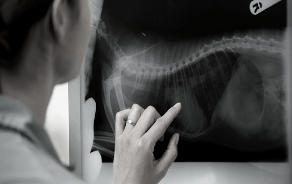
Key Areas
- Develop an effective and practical technique for reading radiographs of dogs and cats
- Practice your radiograph reading skills using a large number of radiographic cases
- Assess your improvement in radiographic interpretation at appropriate intervals through online assessment exercises
About this course
Struggling with knowing whether that white bit on the lung is really a problem? Worried that you might miss an intestinal obstruction of a foreign body? Wondering what is this funky looking lesion in the stifle? In other words – would you like to enhance your confidence in interpreting radiographs?
During this course, you will enhance your knowledge of small animal radiology, create your own radiographic reading technique and sharpen your skills at reading radiographs. A variety of radiographic cases will be used for individual review and group discussions. This will enable you to practice your reading technique and to share your experiences while enhancing your skills in assessing radiographs.
Why do this course?
At the end of this course you will have developed your own structured system of evaluating radiographs, acquired experience in reporting and diagnosing common and more challenging radiographic abnormalities and developed a logical approach to reading and interpreting a radiographic study.
DAY 1 – Radiography of Thorax
| Time |
Title |
| 09:00 – 09:15 |
Introduction |
| 09:15 – 10:45 |
How to read radiographs: the technique |
| 10:45 – 11:30 |
Practice cases: reading technique |
| 11:30 – 12:00 |
Coffee/Tea break |
| 12:00 – 12:30 |
Thoracic radiology: normal radiographic anatomy |
| 12:30 – 13:15 |
Thoracic radiology: lungs |
| 13:15 – 14:00 |
Practice cases: lungs |
| 14:00 – 14:45 |
Lunch |
| 14:45 – 15:30 |
Thoracic radiology: thoracic wall, diaphragm, pleural cavity, mediastinum |
| 15:30 – 16:15 |
Practice cases: thoracic wall, diaphragm, pleural cavity, mediastinum |
| 16:15 – 16:30 |
Coffee/Tea break |
| 16:30 – 17:30 |
Practice cases: Thorax |
| 17:30 |
Closing |
DAY 2 – Abdomen and musculoskeletal radiography
| Time |
Title |
| 09:00 – 09:45 |
Abdominal radiology: normal radiographic anatomy |
| 09:45 – 11:15 |
Abdominal radiology: Liver, spleen, gastrointestinal tract, Urinary tract, genital tracts, peritoneum |
| 11:15 – 11:45 |
Coffee/Tea break |
| 11:45 – 13:30 |
Practice cases: Abdomen |
| 13:30 – 14:30 |
Lunch |
| 14:30 – 16:00 |
Musculoskeletal radiology |
| 16:00 – 16:30 |
Coffee/Tea break |
| 16:30 – 17:30 |
Practice cases: musculoskeletal |
| 17:30 |
Closing |