by tatkonasajt | Oct 9, 2023
DAY 1
| Time |
Title |
Format |
| 09:00 – 09:30 |
Introduction to anesthesia in small animals |
Theoretical |
| 09:30 – 11:00 |
Clinical pharmacology of sedatives, analgesics and hypnotics in veterinary anesthesia |
Theoretical |
| 11:00 – 11:30 |
Coffee break |
|
| 11:30 – 12:30 |
Clinical use of intraoperative monitoring and hemodynamic support |
Theoretical |
| 12:30 – 13:30 |
Breathing systems |
Theoretical |
| 13:30 – 14:30 |
Lunch |
|
| 14:30 – 17:00 |
Loco-regiona anaesthesia part I (Introduction, Sciatic Nerve Block, Saphenous Nerve Block, Testicular Block, LIA, Maxillary Nerve Block, Infraorbital Nerve Block, Caudal alveolar inferior Nerve Block) |
Theoretical |
DAY 2
| Time |
Title |
Format |
| 09:00 – 10:00 |
Cardiovascular complications |
Theoretical |
| 10:00 – 11:00 |
Respiratory and other complications during anesthesia |
Theoretical |
| 11:00 – 11:30 |
Coffee break |
|
| 11:30 – 12:30 |
Ventilation strategies in small animal anesthesia |
Theoretical |
| 12:30 – 13:30 |
Anesthesia of a patient with cardiac diseases |
Theoretical |
| 13:30 – 14:30 |
Lunch |
|
| 14:30 – 17:00 |
Loco-regiona anaesthesia part I (Introduction, Sciatic Nerve Block, Saphenous Nerve Block, Testicular Block, LIA, Maxillary Nerve Block, Infraorbital Nerve Block, Caudal alveolar inferior Nerve Block) |
Practical |
by tatkonasajt | Sep 9, 2023
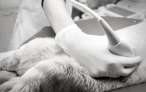
Dvodnevni kurs ultrazvučne dijagnostike koji za cilj ima savladavanje tehnike pregleda nadbubrežnih žlezda, pankreasa, pluća, štitaste žlezde, većih krvnih sudova i važnijih limfnih čvorova. Na kursu će se obraditi i teme brzog pregleda abdomena i grudnog koša (AFAST i TFAST) kao i ultrazvučno vođena punkcija.
Prvi dan
| Vreme |
Naslov |
Format |
| 09:00 – 09:30 |
Uvod |
Teorijski |
| 09:30 – 10:30 |
Tehnika pregleda abdominalnih organa |
Praktično |
| 10:30 – 11:00 |
Pauza za kafu |
|
| 11:00 – 12:00 |
Pankreas, krvni sudovi i važniji limfni čvorovi |
Teorijski |
| 12:00 – 13:30 |
Pankreas, krvni sudovi i važniji limfni čvorovi |
Praktično |
| 13:30 – 14:30 |
Pauza za ručak |
|
| 14:30 – 17:00 |
Pankreas, krvni sudovi i važniji limfni čvorovi |
Praktično |
Drugi dan
| Vreme |
Naslov |
Format |
| 09:00 – 09:30 |
Nadbubrežne žlezde |
Teorijski |
| 09:30 – 10:30 |
Nadbubrežne žlezde |
Praktično |
| 10:30 – 11:00 |
Pauza za kafu |
|
| 11:00 – 11:45 |
Štitasta žlezda, pluća, AFAST, TFAST |
Teorijski |
| 11:45 – 13:30 |
Štitasta žlezda, pluća, AFAST, TFAST |
Praktično |
| 13:30 – 14:30 |
Pauza za ručak |
|
| 14:30 – 16:00 |
Pregled kompletnog abdomena |
Praktično |
| 16:00 – 16:30 |
Ultrazvučno vođena punkcija |
Teorijski |
| 16:30 – 17:00 |
Ultrazvučno vođena punkcija |
Praktično |
by tatkonasajt | Sep 9, 2023
Dvodnevni kurs ultrazvučne dijagnostike abdominalnih organa koji za cilj ima upoznavanje polaznika sa mogućnostima ultrazvučne dijagnostike, savladavanje tehnike pregleda jetre, žučne kese, slezine, bubrega, mokraćne bešike, muškog i ženskog reproduktivnog trakta, digestivnog trakta, desnog režnja pankreasa kao i upoznavanje sa najčešćim ultrazvučno vidljivim patologijama.
Prvi dan
| Vreme |
Naslov |
Format |
| 09:00 – 10:00 |
Uvod i osnove |
Teorijski |
| 10:00 – 10:30 |
Upoznavanje sa aparatom |
Praktično |
| 10:30 – 11:00 |
Pauza za kafu |
|
| 11:00 – 11:45 |
Jetra, žučna kesa, slezina |
Teorijski |
| 11:45 – 13:30 |
Jetra, žučna kesa, slezina |
Praktično |
| 13:30 – 14:30 |
Pauza za ručak |
|
| 14:30 – 15:15 |
Bubrezi, mokraćna bešika |
Teorijski |
| 15:15 – 16:30 |
Bubrezi, mokraćna bešika |
Praktično |
| 16:30 – 17:00 |
Pitanja |
|
Drugi dan
| Vreme |
Naslov |
Format |
| 09:00 – 09:45 |
Želudac, creva, pankreas |
Teorijski |
| 09:45 – 10:30 |
Želudac, creva, pankreas |
Praktično |
| 10:30 – 11:00 |
Pauza za kafu |
|
| 11:00 – 11:30 |
Ženski i muški reproduktivni trakt |
Teorijski |
| 11:30 – 13:30 |
Ženski i muški reproduktivni trakt |
Praktično |
| 13:30 – 14:30 |
Pauza za ručak |
|
| 14:30 – 16:00 |
Praktičan rad |
Praktično |
| 16:00 – 16:30 |
Pisanje izveštaja |
Teorijski |
| 16:30 – 17:00 |
Pitanja |
|

by tatkonasajt | Aug 14, 2023
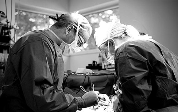
This course combines practical and theoretical parts of head and neck soft tissue surgery. Most of the time will be spent in the wet lab performing surgeries but enough theory will be given so the candidate is able to make good decisions on when and how to do the surgeries but also how to manage complications. The course is suitable for all levels from beginners to advanced as there will be something to be gained by everyone. It is also designed to be flexible so people can practice at their own pace and concentrate on techniques that they would like to perfect. The tutors will be available to observe, and have direct involvement in teaching the procedures.
Learning objectives
- To have an understanding of anatomy of the head and neck and related surgeries
- To understand the application and when to select different surgical procedures of the head and neck
- To learn enough of the theory to enable decision making when deciding which surgeries to perform
- To have knowledge of the risks and complications of each procedure and how to reduce these.
Jackie Demetriou will be one of the course tutors. She is a European Boarded Specialist in Small Animal Surgery, who has practiced specialist level soft tissue surgery for the past 20 years. She has worked in both academia (University of Edinburgh, University of Cambridge and University of Nottingham) and private referral practice. She has extensive experience teaching general practitioners the theory and practice of surgery and really enjoys seeing how surgeons gain confidence in managing cases and performing surgeries after these CPD events.
Day 1
- Ear surgery
- Pinnectomy, biopsy
- Lateral wall resection
- Total ear canal ablation / bulla osteotomy
- Ventral bulla osteotomy (cat)
- Mandibulectomy – rostral and central
- Maxillectomy – rostral and central
- BOAS surgery
Day 2
- Nasal surgery
- Planectomy
- Dorsal rhinotomy
- Neck exploration
- Thyroidectomy
- Sialoadenectomy (mandibular)
- Laryngeal tie back
- tracheostomy
by tatkonasajt | Aug 7, 2023
Objectives and goals of the course
After studying Veterinary Medicine at the Vet School Hannover and a short period in a private practice Dr. Neumann has started working at the Institute of Veterinary Medicine, University of Goettingen in 1992.
In 1996. he received the diploma as a national specialist for small animals and in 1998. for clinical pathology. In 2004. he received the certificate of the ECVCP. 2007. he finished his habilitation and since 2010. he is Professor at the University of Goettingen.
His scientific fields of interest are; orthopedic diseases, especially osteoarthritis; cytokine research of different disease mechanisms and development of biomarkers, gastroenterology, especially liver diseases.
Dr. Neumann is author of about 70 publications in international journals and he is lecturing at four faculties in the fields of small animal surgery, CT, general subjects and clinical pathology.
Dr. Neumann is active in continuing education and specialization in Lower Saxony and nationwide.
DAY 1
| Time |
Contents |
| 09:00 |
Common causes of front and hind limb lameness Shoulder surgery (OCD, Biceps tenotomy, M. infraspinatus myotomy) |
| 10:30 |
Coffee Break |
| 11:00 |
PRACTICAL WORK
Shoulder surgery (OCD, Biceps tenotomy, M. infraspinatus myotomy) |
| 13:00 |
Lunch Break |
| 14:00 |
Elbow surgery (IPC resection, IPA resection, Ulna osteotomy) |
| 15:30 |
Coffee Break |
| 16:00 |
PRACTICAL WORK
Elbow surgery (IPC resection, IPA resection, Ulna osteotomy) |
| 17:30 |
End of the day |
DAY 2
| Time |
Contents |
| 09:00 |
Hip surgery (Symphysiodesis, Joint capsule suturing, Denervation, Femoral head and neck resection) |
| 10:30 |
Coffee Break |
| 11:00 |
PRACTICAL WORK
Hip surgery (Symphysiodesis, Joint capsule suturing, Denervation, Femoral head and neck resection) |
| 13:00 |
Lunch Break |
| 14:00 |
Stifle surgery (Ruptured cranial cruciate ligament, patella luxation) |
| 15:30 |
Coffee Break |
| 16:00 |
PRACTICAL WORK
Stifle surgery (Ruptured cranial cruciate ligament, patella luxation) |
| 17:30 |
End of the course |
