by tatkonasajt | Dec 26, 2023
Ključne oblasti
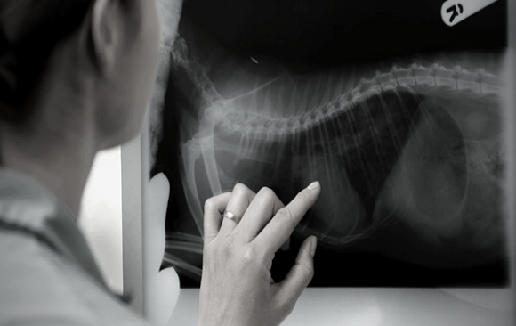
- Mučite se da saznate da li je taj beli deo na plućima zaista patologija?
- Brinete da biste mogli da propustite crevnu opstrukciju usled stranog tela?
- Niste sigurni koje su radiografske indikacije za hiruršku intervenciju!
Šta vam omogućava ovaj kurs?
- Razvijanje efikasne i praktične tehnike za čitanje radiografskih snimaka pasa i mačaka
- Praktičan rad u grupama koristeći veliki broj radiografskih slučajeva uz klinički pristup
Tokom ovog kursa ćete poboljšati svoje znanje iz radiografije pasa i mačaka, kreirati sopstvenu tehniku čitanja radiografskih snimaka i izoštriti svoje kliničke veštine.
Rad u malim grupama će vam omogućiti da uvežbate svoju tehniku čitanja i da podelite svoja iskustva sa kolegama.
Nadamo se da ćete na kraju ovog kursa razviti sistematični pristup proceni snimaka, steći iskustvo u dijagnostikovanju zastupljenijih, ali i ređih radiografskih abnormalnosti i razviti logičan pristup tumačenju radiografske studije.
DAN 1 – Radiologija toraksa
- PREDAVANJE : Generalni principi I tehnički aspekti radiologije toraksa
- PRAKTIČNI : Radiološko pozicioniranje I podešavanja ekspozicije
- PREDAVANJE : Tumačenje bolesti pluća
- RADIONICA: Čitanje snimaka
- PREDAVANJE: Radiološko tumačenje kardioloških bolesti
- PREDAVANJE: Pleura, medijastinum, dijafragma i zid toraksa
- RADIONICA: Čitanje snimaka
DAN 2 – Radiologija abdomena
- PREDAVANJE: Generalni principi I tehnički aspekti radiologije abdomena
- PRAKTIČNI: Radiološko pozicioniranje I podešavanja ekspozicije
- PREDAVANJE: Koje se korisne kliničke informacije mogu dobiti na snimcima abdomena?
Otkrivanje abdominalnih masa, tečnosti i slobodnog gasa.
Znaci gastrointestinalne opstrukcije.
Znaci bolesti urinarnog trakta.
- RADIONICA: Čitanje snimaka
- PREDAVANJE: Gastrointestinalne i genitourinarne kontrasne studije.
Kada, zašto I kako bi to trebali da radite?
- RADIONICA: Čitanje snimaka
by tatkonasajt | Dec 22, 2023
This course is approved by GRSK and will cover the following subjects:
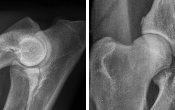
- Introduction to hip dysplasia and elbow dysplasia.
- Possibilities to reduce dysplasia: how can breeding influence the prevalence of HD and ED.
- Radiographic technique – practical on how to take a proper radiographs for the assessment of the hip and elbow dysplasia. Quality evaluation of the radiographs for the HD and ED.
- Explanation of different scoring systems.
- Film reading session – scoring of the HD ED radiographs.
DAY1
| Time |
Title |
| 09.00 – 09.15 |
Welcome and Introduction |
| 09.15 – 10.30 |
Introduction to hip dysplasia and elbow dysplasia
Possibilities to reduce dysplasia
How can breeding influence the prevalence of HD and ED |
| 10.30 – 11.00 |
Coffee break |
| 11.00 – 12.00 |
How to take a proper radiograph for the HD assessment |
| 12.00 – 13.00 |
How to take a proper radiograph for the ED assessment |
| 13.15 – 14.30 |
Lunch break |
| 14.30 – 16.30 |
Radiographic technique – practical |
| 16.30 – 17.00 |
Coffee break |
| 17.00 – 18.30 |
Film reading session on radiographic film quality |
DAY2
| Time |
Title |
| 09.00 – 10.30 |
HD scoring systems / protocols
Radiographic findings and evaluation of HD
Scoring criteria and grading of HD |
| 10.30 – 11.00 |
Coffee break |
| 11.00 – 12.30 |
HD film reading individual work and group work |
| 12.30 – 13.30 |
F.C.I. – Kennel Clubs, Breed Clubs – HD ED control |
| 13.30 – 14.30 |
Lunch break |
| 14.30 – 16.00 |
ED scoring system / protocol
Radiographic findings and evaluation of ED
Scoring criteria and grading of ED |
| 16.00 – 16.30 |
Coffee break |
| 16.30 – 18.00 |
ED film reading individual work and group work |
| 18.00 – 18.30 |
Closing remarks |
by tatkonasajt | Aug 7, 2023
This course is approved by GRSK and will cover the following subjects:
- Introduction to hip dysplasia and elbow dysplasia.
- Possibilities to reduce dysplasia: how can breeding influence the prevalence of HD and ED.
- Radiographic technique – practical on how to take a proper radiographs for the assessment of the hip and elbow dysplasia. Quality evaluation of the radiographs for the HD and ED.
- Explanation of different scoring systems.
- Film reading session – scoring of the HD ED radiographs.
DAY1
| Time |
Title |
| 09.00 – 09.15 |
Welcome and Introduction |
| 09.15 – 10.30 |
Introduction to hip dysplasia and elbow dysplasia
Possibilities to reduce dysplasia
How can breeding influence the prevalence of HD and ED |
| 10.30 – 11.00 |
Coffee break |
| 11.00 – 12.00 |
How to take a proper radiograph for the HD assessment |
| 12.00 – 13.00 |
How to take a proper radiograph for the ED assessment |
| 13.15 – 14.30 |
Lunch break |
| 14.30 – 16.30 |
Radiographic technique – practical |
| 16.30 – 17.00 |
Coffee break |
| 17.00 – 18.30 |
Film reading session on radiographic film quality |
DAY2
| Time |
Title |
| 09.00 – 10.30 |
HD scoring systems / protocols
Radiographic findings and evaluation of HD
Scoring criteria and grading of HD |
| 10.30 – 11.00 |
Coffee break |
| 11.00 – 12.30 |
HD film reading individual work and group work |
| 12.30 – 13.30 |
F.C.I. – Kennel Clubs, Breed Clubs – HD ED control |
| 13.30 – 14.30 |
Lunch break |
| 14.30 – 16.00 |
ED scoring system / protocol
Radiographic findings and evaluation of ED
Scoring criteria and grading of ED |
| 16.00 – 16.30 |
Coffee break |
| 16.30 – 18.00 |
ED film reading individual work and group work |
| 18.00 – 18.30 |
Closing remarks |
by tatkonasajt | Mar 19, 2023
Key Areas
- Develop an effective and practical technique for reading radiographs of dogs and cats
- Practice your radiograph reading skills using a large number of radiographic cases
- Assess your improvement in radiographic interpretation at appropriate intervals through online assessment exercises
About this course
Struggling with knowing whether that white bit on the lung is really a problem? Worried that you might miss an intestinal obstruction of a foreign body? Wondering what is this funky looking lesion in the stifle? In other words – would you like to enhance your confidence in interpreting radiographs?
During this course, you will enhance your knowledge of small animal radiology, create your own radiographic reading technique and sharpen your skills at reading radiographs. A variety of radiographic cases will be used for individual review and group discussions. This will enable you to practice your reading technique and to share your experiences while enhancing your skills in assessing radiographs.
Why do this course?
At the end of this course you will have developed your own structured system of evaluating radiographs, acquired experience in reporting and diagnosing common and more challenging radiographic abnormalities and developed a logical approach to reading and interpreting a radiographic study.
DAY 1 – Radiography of Thorax
| Time |
Title |
| 09:00 – 09:15 |
Introduction |
| 09:15 – 10:45 |
How to read radiographs: the technique |
| 10:45 – 11:30 |
Practice cases: reading technique |
| 11:30 – 12:00 |
Coffee/Tea break |
| 12:00 – 12:30 |
Thoracic radiology: normal radiographic anatomy |
| 12:30 – 13:15 |
Thoracic radiology: lungs |
| 13:15 – 14:00 |
Practice cases: lungs |
| 14:00 – 14:45 |
Lunch |
| 14:45 – 15:30 |
Thoracic radiology: thoracic wall, diaphragm, pleural cavity, mediastinum |
| 15:30 – 16:15 |
Practice cases: thoracic wall, diaphragm, pleural cavity, mediastinum |
| 16:15 – 16:30 |
Coffee/Tea break |
| 16:30 – 17:30 |
Practice cases: Thorax |
| 17:30 |
Closing |
DAY 2 – Abdomen and musculoskeletal radiography
| Time |
Title |
| 09:00 – 09:45 |
Abdominal radiology: normal radiographic anatomy |
| 09:45 – 11:15 |
Abdominal radiology: Liver, spleen, gastrointestinal tract, Urinary tract, genital tracts, peritoneum |
| 11:15 – 11:45 |
Coffee/Tea break |
| 11:45 – 13:30 |
Practice cases: Abdomen |
| 13:30 – 14:30 |
Lunch |
| 14:30 – 16:00 |
Musculoskeletal radiology |
| 16:00 – 16:30 |
Coffee/Tea break |
| 16:30 – 17:30 |
Practice cases: musculoskeletal |
| 17:30 |
Closing |
by tatkonasajt | Jan 2, 2023
This course is approved by GRSK and will cover the following subjects:
- Introduction to hip dysplasia and elbow dysplasia.
- Possibilities to reduce dysplasia: how can breeding influence the prevalence of HD and ED.
- Radiographic technique – practical on how to take a proper radiographs for the assessment of the hip and elbow dysplasia. Quality evaluation of the radiographs for the HD and ED.
- Explanation of different scoring systems.
- Film reading session – scoring of the HD ED radiographs.
DAY1
| Time |
Title |
| 09.00 – 09.15 |
Welcome and Introduction |
| 09.15 – 10.30 |
Introduction to hip dysplasia and elbow dysplasia
Possibilities to reduce dysplasia
How can breeding influence the prevalence of HD and ED |
| 10.30 – 11.00 |
Coffee break |
| 11.00 – 12.00 |
How to take a proper radiograph for the HD assessment |
| 12.00 – 13.00 |
How to take a proper radiograph for the ED assessment |
| 13.15 – 14.30 |
Lunch break |
| 14.30 – 16.30 |
Radiographic technique – practical |
| 16.30 – 17.00 |
Coffee break |
| 17.00 – 18.30 |
Film reading session on radiographic film quality |
DAY2
| Time |
Title |
| 09.00 – 10.30 |
HD scoring systems / protocols
Radiographic findings and evaluation of HD
Scoring criteria and grading of HD |
| 10.30 – 11.00 |
Coffee break |
| 11.00 – 12.30 |
HD film reading individual work and group work |
| 12.30 – 13.30 |
F.C.I. – Kennel Clubs, Breed Clubs – HD ED control |
| 13.30 – 14.30 |
Lunch break |
| 14.30 – 16.00 |
ED scoring system / protocol
Radiographic findings and evaluation of ED
Scoring criteria and grading of ED |
| 16.00 – 16.30 |
Coffee break |
| 16.30 – 18.00 |
ED film reading individual work and group work |
| 18.00 – 18.30 |
Closing remarks |