by tatkonasajt | Dec 22, 2023
After studying Veterinary Medicine at the Vet School Hannover and a short period in a private practice Dr. Neumann has started working at the Institute of Veterinary Medicine, University of Goettingen in 1992.
In 1996. he received the diploma as a national specialist for small animals and in 1998. for clinical pathology. In 2004. he received the certificate of the ECVCP. 2007. he finished his habilitation and since 2010. he is Professor at the University of Goettingen.
His scientific fields of interest are; orthopedic diseases, especially osteoarthritis; cytokine research of different disease mechanisms and development of biomarkers, gastroenterology, especially liver diseases.
Dr. Neumann is author of about 70 publications in international journals and he is lecturing at four faculties in the fields of small animal surgery, CT, general subjects and clinical pathology.
Dr. Neumann is active in continuing education and specialization in Lower Saxony and nationwide.
DAY 1
| Time |
Title |
| 09:00 – 10:30 |
Common causes of front and hind limb lameness Shoulder surgery (OCD, Biceps tenotomy, M. infraspinatus myotomy) |
| 10:30 – 11:00 |
Coffee break |
| 11:00 – 13:00 |
PRACTICAL WORK: Shoulder surgery (OCD, Biceps tenotomy, M. infraspinatus myotomy) |
| 13:00 – 14:00 |
Lunch break |
| 14:00 – 15:30 |
Elbow surgery (IPC resection, IPA resection, Ulna osteotomy) |
| 15:30 – 16:00 |
Coffee break |
| 16:00 – 17:30 |
PRACTICAL WORK: Elbow surgery (IPC resection, IPA resection, Ulna osteotomy) |
DAY 2
| Time |
Title |
| 09:00 – 10:30 |
Hip surgery (Symphysiodesis, Joint capsule suturing, Denervation, Femoral head and neck resection) |
| 10:30 – 11:00 |
Coffee break |
| 11:00 – 13:00 |
PRACTICAL WORK: Hip surgery (Symphysiodesis, Joint capsule suturing, Denervation, Femoral head and neck resection) |
| 13:00 – 14:00 |
Lunch break |
| 14:00 – 15:30 |
Stifle surgery (Ruptured cranial cruciate ligament, patella luxation) |
| 15:30 – 16:00 |
Coffee break |
| 16:00 – 17:30 |
PRACTICAL WORK: Stifle surgery (Ruptured cranial cruciate ligament, patella luxation) |
by tatkonasajt | Aug 7, 2023
Objectives and goals of the course
After studying Veterinary Medicine at the Vet School Hannover and a short period in a private practice Dr. Neumann has started working at the Institute of Veterinary Medicine, University of Goettingen in 1992.
In 1996. he received the diploma as a national specialist for small animals and in 1998. for clinical pathology. In 2004. he received the certificate of the ECVCP. 2007. he finished his habilitation and since 2010. he is Professor at the University of Goettingen.
His scientific fields of interest are; orthopedic diseases, especially osteoarthritis; cytokine research of different disease mechanisms and development of biomarkers, gastroenterology, especially liver diseases.
Dr. Neumann is author of about 70 publications in international journals and he is lecturing at four faculties in the fields of small animal surgery, CT, general subjects and clinical pathology.
Dr. Neumann is active in continuing education and specialization in Lower Saxony and nationwide.
DAY 1
| Time |
Contents |
| 09:00 |
Common causes of front and hind limb lameness Shoulder surgery (OCD, Biceps tenotomy, M. infraspinatus myotomy) |
| 10:30 |
Coffee Break |
| 11:00 |
PRACTICAL WORK
Shoulder surgery (OCD, Biceps tenotomy, M. infraspinatus myotomy) |
| 13:00 |
Lunch Break |
| 14:00 |
Elbow surgery (IPC resection, IPA resection, Ulna osteotomy) |
| 15:30 |
Coffee Break |
| 16:00 |
PRACTICAL WORK
Elbow surgery (IPC resection, IPA resection, Ulna osteotomy) |
| 17:30 |
End of the day |
DAY 2
| Time |
Contents |
| 09:00 |
Hip surgery (Symphysiodesis, Joint capsule suturing, Denervation, Femoral head and neck resection) |
| 10:30 |
Coffee Break |
| 11:00 |
PRACTICAL WORK
Hip surgery (Symphysiodesis, Joint capsule suturing, Denervation, Femoral head and neck resection) |
| 13:00 |
Lunch Break |
| 14:00 |
Stifle surgery (Ruptured cranial cruciate ligament, patella luxation) |
| 15:30 |
Coffee Break |
| 16:00 |
PRACTICAL WORK
Stifle surgery (Ruptured cranial cruciate ligament, patella luxation) |
| 17:30 |
End of the course |
by tatkonasajt | Mar 26, 2023
Objectives and goals of the course
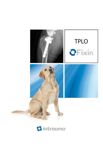
This TPLO course is designed for practicing veterinarians with experience in small animal orthopaedic surgery. The participants will became familiar with the planning and execution of the most commonly performed procedure in cranial cruciate deficient stifle using one of the most versatile TPLO locking system.
The detailed theoretical sessions will bring the knowledge of rationale, case selection, preoperative planning, surgical execution and potential complications in TPLO procedure. The structure of the labs will allows the participants to learn the procedure in a “step by step” manner first on a bone models and then on a cadaver species.
DAY 1
| Time |
Title |
| 08:30 – 09:00 |
Understanding stifle biomechanics and rationale behind TPLO |
| 09:00 – 09:30 |
Radiographic positioning and TPA measuring |
| 09:30 – 10:00 |
TPLO implants and equipment |
| 10:00 – 10:30 |
Coffee break |
| 10:30 – 11:00 |
Preoperative planning of TPLO |
| 11:00 – 12:30 |
TPLO – step by step description of the procedure. Postoperative radiographic assessment after TPLO |
| 12:30 – 13:30 |
Lunch break |
| 13:30 – 14:00 |
Complications related to the TPLO procedure |
| 14:00 – 15:00 |
Preoperative planning of TPLO – practical exercise (drawing on a printed radiographs, TPA calculation, defining the best position of the cut, implant choice and position) – TPLO charts, saw blade radius templates and plates templates will be needed for this exercise |
| 15:00 – 17:00 |
Getting familiar with the equipment and performing TPLO on a bone model – laboratory |
| 17:00 – 17:30 |
Summary and question session and end of the course |
DAY 2
| Time |
Title |
| 08:30 – 10:00 |
Preoperative planning on x-rayed cadaver limbs |
| 10:00 – 10:30 |
Coffee break |
| 10:30 – 12:00 |
TPLO on a cadaver limb – practical session 1 |
| 12:00 – 13:00 |
Lunch break |
| 13:00 – 14:30 |
TPLO on a cadaver limb – practical session 2 |
| 14:30 – 15:30 |
Discussion of postoperative radiographs |
| 15:30 – 16:00 |
What’s the difference in Giant breeds |
| 16:00 – 16:30 |
What’s the difference in small dogs. TPLO + patella luxation |
| 16:30 – 17:00 |
Summary, question session and end of the course |
by tatkonasajt | Mar 26, 2023
Objectives and goals of the course
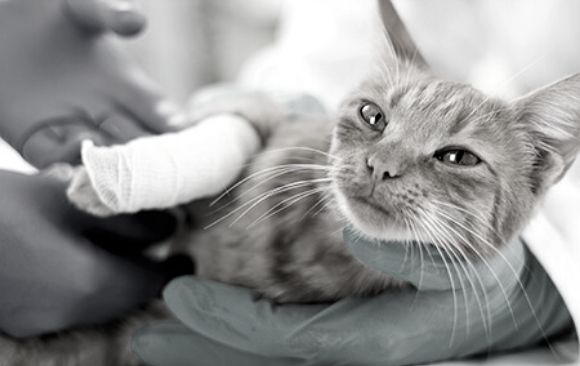
This two days course is designed for veterinary practitioners with some experience in treatment of fractures and other orthopaedic conditions in small animals. Some anatomical differences makes the orthopaedic care in feline patients more challenging and might reflects to the treatment options. The course will consists of theoretical sessions alternating with small group practicals on cadaver specimens. This will allows the participants to implement the knowledge delivered during the theoretical parts in the practical sessions starting with performing the surgical approach to different bones and using different implants for stabilising some of the most commonly seen fractures in feline patients.
Overview of most common fracture scenarios in feline patients
Decision making process in fracture planning
Interactive case-oriented discussion
After the course, participants will be able to construct a treatment plan and will become familiar how to treat most common orthopaedic conditions and fractures in feline patients.
DAY 1
| Time |
Title |
| 09:00 |
Welcome |
| 09:15 – 10:15 |
Principles of fracture repair in cats – decision making and treatment options |
| 10:15 – 11:15 |
Common fractures of the front limb |
| 11:15 – 11:30 |
Coffee break |
| 11:30 – 12:30 |
Common fractures of the hind limb |
| 12:30 – 13:30 |
Lunch break |
| 13:30 – 14:30 |
Fractures of the pelvis. Sacral and sacro-coccygeal fracture/luxation |
| 14:30 – 16:00 |
Practical exercise 1 – Application of an orthopaedic plate to a short oblique tibial fracture |
| 16:00 – 17:30 |
Practical exercise 2 – Application of an orthopaedic plate to a comminuted humerus fracture |
| 17:30 – 18:00 |
Questions and summary of day 1 |
DAY 2
| Time |
Title |
| 08:30 – 09:30 |
Traumatic joint luxations in cats |
| 09:30 – 10:00 |
Cat with patella luxation |
| 10:00 – 10:30 |
Cat with cruciate ligament deficient stifle |
| 10:30 – 11:00 |
Coffee break |
| 11:00 – 12:00 |
Head trauma. Mandibular and maxillary fractures |
| 12:00 – 13:00 |
Practical exercise 3 – Cross pin fixation in Salter Harris fracture of the distal femoral growth plate |
| 13:00 – 14:00 |
Lunch break |
| 14:00 – 15:00 |
Practical exercise 4 – Approach to the hip joint and iliofemoral suture application |
| 15:00 – 16:00 |
Dorsal approach to the hip joint and application of plate craniodorsal to the acetabulum |
| 16:00 – 17:00 |
“If you put a cat and a bunch of brocken bones in the same room, the bones will heal! “ Challenging fracture cases in cats – interactive discussion |
| 17:00 – 17:30 |
Questions, summary and end of the course |