Neurology 1
HOW TO DESCRIBE WHAT YOU SEE AND KNOW IF IT IS NORMAL
The neurological examination is one of the most important and cost-effective tools in clinical neurology
During this course delegate will learn:
- To determine if the nervous system is affected in a disease process (to detect the presence of a neurological abnormality)
- To establish an accurate anatomical diagnosis, to localise the lesion (to determine its location)
The neurological examination is the most important step in identifying if a patient’s condition is neurological (versus medical or orthopedic) and where in the nervous system the pathology is located. Delegate will be explained how to establish a routine standard procedure for examining an animal. This will provide the experience and confidence necessary to make an accurate neuroanatomical diagnosis. The neurological examination should enable an anatomical diagnosis to be established and determine where the lesion is: Brain, Spinal cord or Peripheral nerve and muscle. It is important to consider if one lesion can explain all deficits, or if the disease is more diffuse or multifocal.
This course is aimed at the every clinician as well to those with more experience and interest in small animal neurology.
DAY 1
| Time | Title |
| 09:00 – 09:15 | Welcome and Introduction |
| 09:15 – 09:45 | Multiple Choice Test |
| 09:45 – 11:00 | Talk: Neurological Examination |
| 11:00 – 11:30 | Coffee break |
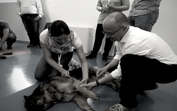
| 11:30 – 12:00 | Demonstration of Neurological examination |
| 12:00 – 13:15 | Neurological examination performed by participants |
| 13:15 – 14:15 | Lunch |
| 14:15 – 15:45 | Talk: Neurolocalisation |
| 15:45 – 16:15 | Coffee break |
| 16:15 – 17:45 | Neurolocalisation with everybody together based on videos Part I (6 cases) |
DAY 2
| Time | Title |
| 09:00 – 11:00 | Neurolocalisation based on videos in groups part II (6 cases) |
| 11:00 – 11:30 | Coffee break |
| 11:30 – 13:00 | Cases from the first session of the day are presented by the participants to everybody and discussed together |
| 13:00 – 14:00 | Lunch |
| 14:00 – 15:45 | Neurolocalisations on real patients (if available) |
| 15:45 – 16:15 | Coffee break |
| 16:15 – 17:30 | Discussion of own cases (every participant has to bring one own case) |
| 17:30 – 18:00 | MC-Test to allow the participants to judge the progress they have made during the seminar |